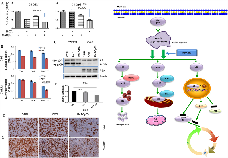
Figure 5.
ReACp53 increases sensitivity of PCa cells receiving treatments of ADT or ENZA. A. Graphs showing the effects of combination treatment of ReACp53 and ENZA on clonogenic survival of C4–2 cells with engineering expression of mutant p53; B. Graphs showing the effects of ReACp53 on cell vialbility of PCa cells receiving ADT; C. Western blot results showing the effect of ReACp53 on expressions of AR and PSA in PCa cells; D. Representative images of IHC staining showing the effect of ReACp53 treatment on expressions of AR protein in xenografts established from CWRR1 or C4–2 cells; E. Graphs showing the changes of PSA transcripts in C4–2 cells treated with 10 μM of ReACp53 for 48 hours. Data represent the average of three independent experiments. Error bars represent standard deviation. * indicates p<0.05; *** indicates p<0.001; NS represents no statistic significance; F. Model for the action of ReACp53 on cell death and cell proliferation of PCa cells carrying mutant p53 protein. ReACp53 treatment segregates the amyloid aggregation of mutant p53 protein complex and induces binding of Mdm2 and Bax to p53 protein, resulting in the degradation of p53 protein and the translocation of p53-Bax to mitochondria to induce mitochondrial cell death in PCa cells, or restores p53 nuclear function as a transcription factor.

