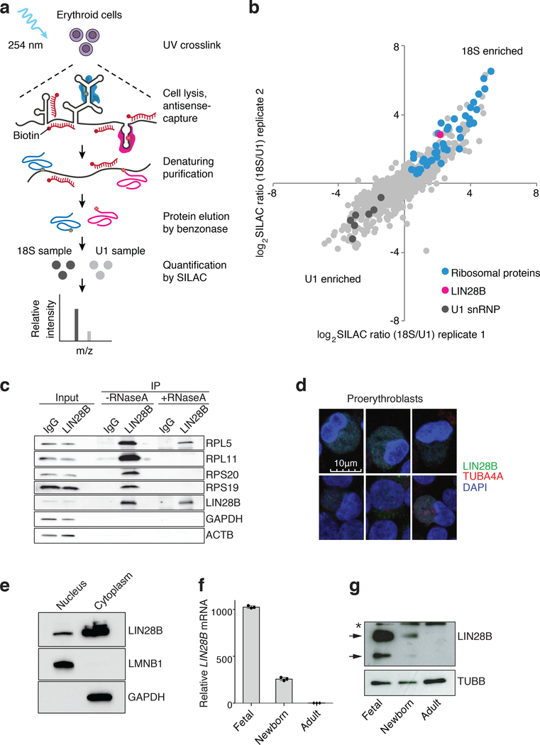
Fig. 2. The RNA binding protein LIN28B associates with ribosomes in erythroid cells and is developmentally regulated.
a, Schematic overview of RAP-MS combined with SILAC mass spectrometry. b, Quantification of 18S and U1 interacting proteins. Scatter plot of log2 transformed SILAC ratios from two biological replicates is shown. c, Immunoprecipitation (IP) in erythroid cells using antibodies targeting LIN28B or control IgG. Western blot detection of the ribosomal proteins RPL5, RPL11, RPS20, RPS19, and LIN28B, with GAPDH and β-actin as controls. -RNaseA and +RNaseA denote IP without or with RNaseA treatment for 30 minutes, respectively. Please note gaps in the western blot between experimental conditions that were placed to ensure appropriate migration patterns for all proteins. Experiment repeated 2 times independently. d, Representative confocal immunocytochemistry images of 4% paraformaldehyde-fixed newborn erythroid cells at differentiation day 7. The nuclear stain is DAPI (blue). LIN28B is detected with Alexa Fluor 488 (green) and tubulin (TUBA4A) with Alexa Fluor 594 (red). Scale bar is shown. Experiment repeated 5 times independently. e, Representative western blots showing subcellular localization of LIN28B, Lamin B1 and GAPDH. The nuclear and cytoplasmic fractions are labeled. Experiment repeated 3 times independently. f, LIN28B mRNA distribution (normalized to GAPDH expression) in fetal, newborn, and adult erythroid cells at differentiation day 7 from HSPCs (n = 3 per time point; 3 biologically independent experiments). Mean is plotted and error bars show s.d. g, Representative western blots showing LIN28B expression in fetal, newborn, and adult at differentiation day 7 from HSPCs (3 independent experiments). Arrows indicate two LIN28B isoforms and asterisk indicates a non-specific band. β-tubulin is used as a loading control. Blots have been cropped and the corresponding full blots are available in the Source Data files.

