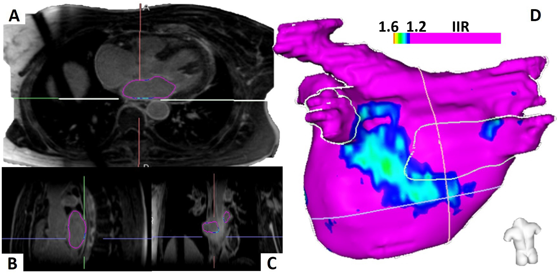
Figure 1.

Left Atrial Segmentation and Three-Dimensional Left Atrium Model. The left atrium is manually contoured on three-dimensional, high-resolution late gadolinium enhancement cardiac magnetic resonance images and visualized in all three axes (A-C). The left atrial three-dimensional model with posterior wall substrate in an ablation-naive patient is displayed (D).
