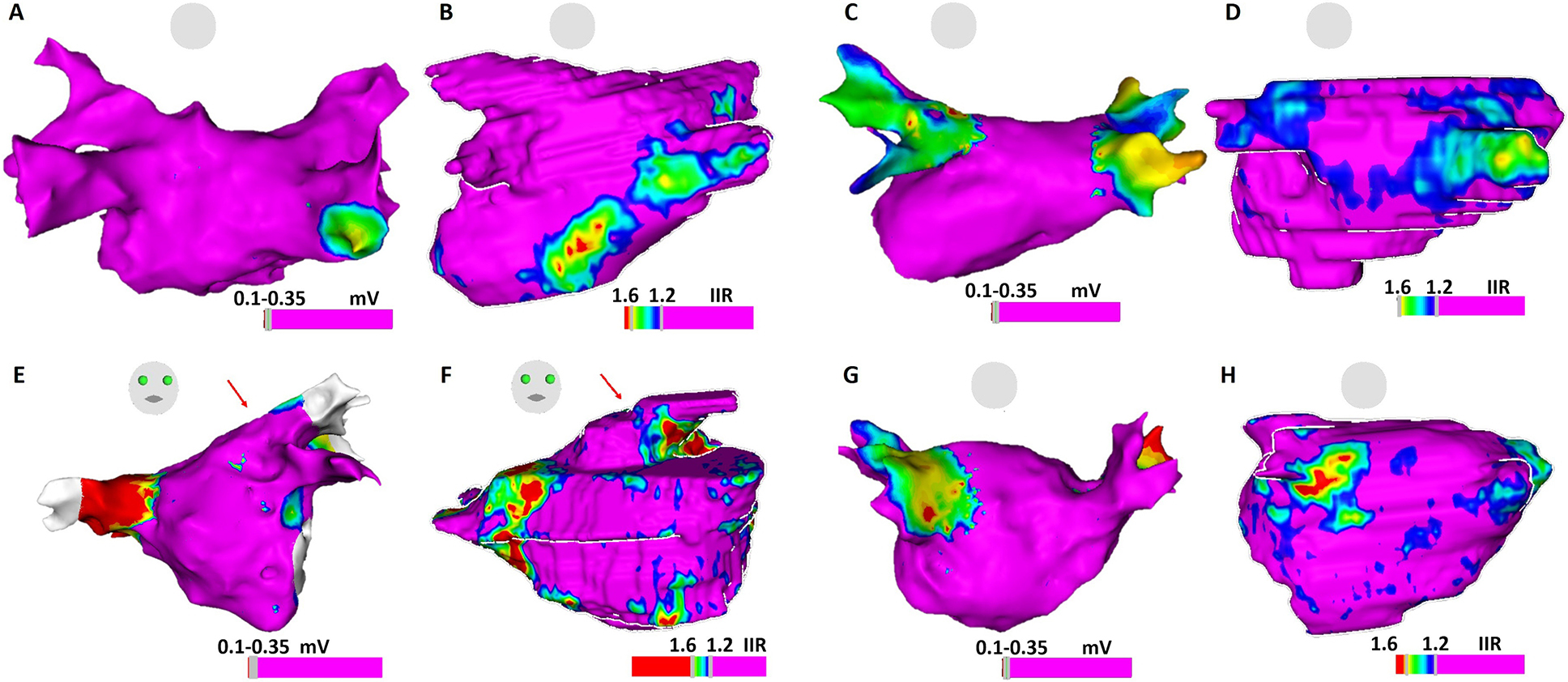
Figure 5.

Comparison of atrial substrate on EAM voltage map and LGE-CMR shell. Panels 5A and 5B: ablation-naïve patient with normal LA voltage but increased EGM fractionation on posterior LGE region on CMR shell; Panels 5C and 5D: Re-do ablation patient with chronic isolated right PVs and reconnected left PVs, compatible with low voltage at the antrum of right PVs on EAM and abnormal atrial myocardium on LGE-CMR; Panels 5E and 5F: Re-do ablation patient with normal voltage, but delayed atrial EGMs (arrow) on left PV antrum, corresponding to the atrial substrate (arrow) on LGE-CMR; Panels 5G and 5H: Electrical reconnection of right PVs. EGMs shows normal voltage at right PV antrum. Few EGMs show delayed activation at scarce atrial substrate region on CMR.
