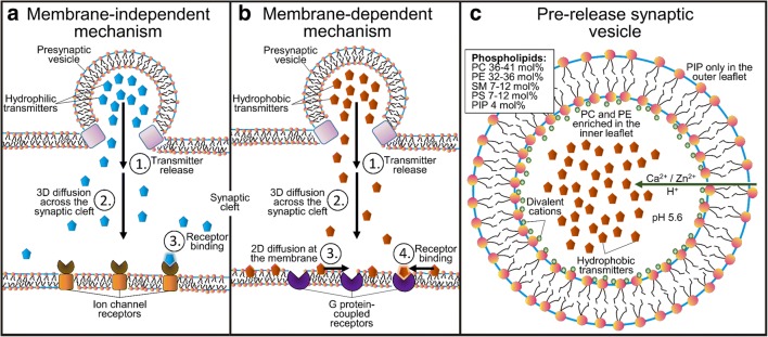
Fig. 5.
Synaptic neurotransmission models. Left panel—membrane-dependent model: (1) release of lipophilic neurotransmitters (NTs), (2) diffusion across the synaptic cleft, (3) binding onto the postsynaptic membrane surface and 2D diffusion on the membrane plane, and, finally, binding into the receptors. Middle panel—membrane-independent model: (1) release of lipophobic NTs, (2) diffusion across the synaptic cleft, and binding into the receptors. Right panel—the presynaptic vesicle with its known lipid composition [77, 78]. Reproduced with the permission from ref. [58]. Copyright 2017 American Chemical Society

