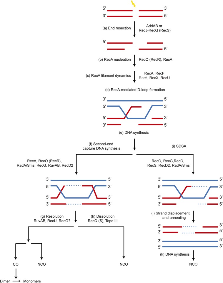
FIGURE 1.
Mechanism of recombinational repair of DSBs in B. subtilis. The main players in each step are shown. Since the SsbA protein influences all processes involving ssDNA, it is not depicted for clarity. (a) the 5′-ends are resected to produce a 3′-tailed duplex by AddAB or by RecJ in concert with a RecQ-like enzyme (RecQ or RecS). SsbA, which interacts with RecJ, RecQ and RecS, binds to the ssDNA region. (b) A set of accessory proteins (RecA mediators, RecO and RecR) act before homology search and contribute to load RecA onto ssDNA displacing bound SsbA, which interacts with RecO. (c) The accessory proteins, known as modulators (RecF, RecX, RecU and RarA [this work]) act during homology search and DNA strand exchange, regulating the formation of dynamic RecA filaments (SsbA interacts with RarA, and RecA with RecU and RecX). (d,e) Strand invasion of the 3′-end of the invading strand results in a D-loop recombination intermediate and provides the primer for DNA synthesis. (f) A capture of the second DNA derived from the other end of the DSB by RecA, RecO, RecR, leads to a double HJ that can be processed by branch migration translocases RecG, RuvAB, RadA/Sms, and may be RecD2. (g) RuvAB in concert with RecU resolves the HJ, leading to CO or NCO products. The CO products lead to dimers that are resolved to monomers by specialized site-specific recombinases. (h) A type I Topoisomerase in concert with a RecQ-like helicase (RecQ, RecS) may dissolve the double HJ, producing only NCO products. (i–k) A synthesis dependent strand annealing mechanism (SDSA) is depicted. The 3′-invading end is extended by the replicase. Then it may be displaced from the joint molecule by branch migration translocases (RuvAB, RecG, RadA/Sms, RecD2, RecQ, RecS) and re-annealed with the complementary strand of the other resected end of the break by the strand annealing activity of RecO, leading to NCO products.

