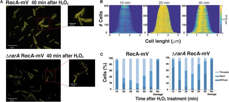
FIGURE 6.
Epifluorescence microscopy showing that RecA assembly into threads is dependent on RarA. (A) Subcellular localization of RecA-mV 40 min after treatment with 0.5 mM H2O2, in wt (rec+) and in ΔrarA mutant cells. Scale bars 5 μm. (B) Demographs of wt B. subtilis cells, demonstrating the localization of RecA-mV to the central regions. Cells were aligned and ordered according to size. The fluorescence profiles represent the mean fluorescence values along the medial axis after background subtraction and normalization such that the maximum fluorescence of each cell is equal. (C) Quantitative analysis of RecA thread formation in wt or rarA mutant cells. The results are the average of three independent experiments (n = 450 cells).

