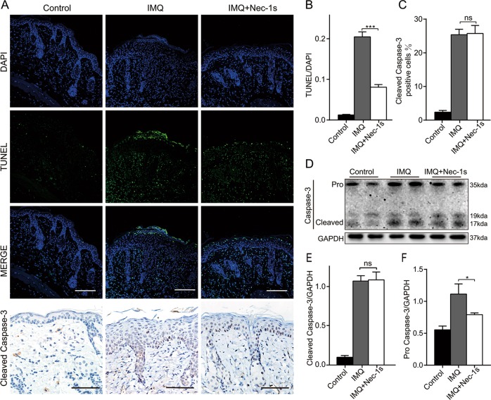
Fig. 5. Nec-1s affected programmed necrosis rather than apoptosis in IMQ-induced psoriasiform dermatitis in mice.
a TUNEL-stained (green fluorescence) cells in the skin biopsies from mice, co-stained with DAPI (blue fluorescence) to detect the nucleus. Immunohistochemical staining for cleaved caspase-3 in the dorsal mouse skin samples of the three groups. Representative images are shown. All scale bars represent 200 μm. TUNEL and cleaved caspase-3-positive cells were quantified under four randomly optical fields per section and normalized over the visual area counted. Four measurements were performed on each field. b The ratio of microscopic quantification of TUNEL-positive cells in three groups. c The ratio of microscopic quantification of cleaved caspase-3-positive cells in epidermis of three groups. d Protein level of caspase-3 was analyzed by western blotting in the three groups of mice. GAPDH served as the loading control. e, f Expression ratio of cleaved caspase-3 and pro caspase 3 to GAPDH was calculated from the relative optical density. Results are representative of three independent experiments. Error bars represent mean ± standard deviation (SD). ns p > 0.05, *p < 0.05, and ***p < 0.001 when compared.

