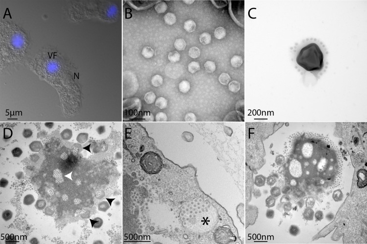
Fig. 1. Microscopy images of MegavirusC vitis and its associated virophage Z. vitis.
a Fluorescence image of DAPI-stained A. castellanii cells infected by MC. vitis and its virophage. Viral particles are visible in the periphery of the viral factory (VF). The cell nucleus (N) remains visible but its fluorescence becomes undetectable due to the intense labelling of the VF DNA. b Transmission electron microscopy (TEM) of Zamilon vitis particles observed by negative staining electron microscopy; c TEM of virophage particles stuck to the giant virus particle (negative staining); d ultrathin section TEM of a MC. vitis viral factory observed in late infection of A. castellanii cells: virophage particles can be seen in holes in the VF (white arrowhead) as well as penetrating a maturing MC. vitis particle (black arrowheads); e neo-synthesized Z. vitis virophage particles gathered in vacuoles (black star) are seen at the periphery of the infected cell suggesting that they are released by exocytosis. f TEM image of an isolated viral factory observed in an ultrathin section of a late infection of A. castellanii cells: virophages accumulate at one pole of the VF as well as in holes in the VF while immature and mature MC. vitis particles are seen at the opposite pole of the VF (Supplementary movie).

