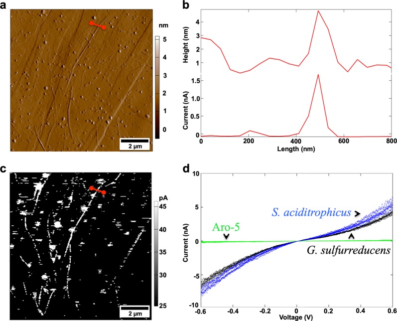
Fig. 2. Characterization of Syntrophus aciditrophicus pili with conductive tip atomic force microscopy.
a Contact mode topographic imaging of pili on highly ordered pyrolytic (HOPG). Red line designates the cross-section examined in b. b Topographic analysis of the height/diameter of an individual pilus and corresponding current measurements (100 mV differential between the tip and the HOPG) across the pilus cross-section. c Current response of the pili shown in a with an applied 100 mV differential. d Current–voltage analysis of individual pili of S. aciditrophicus (blue data points), wild-type G. sulfurreducens (black data points), and the Aro-5 strain of G. sulfurreducens (green data points). Current–voltage spectroscopy is shown for one pilus of each type and is representative of analysis of three distinct locations on three separate pili of each type. Additional scans of S. aciditrophicus pili available in Supplementary Fig. 2. The G. sulfurreducens wild-type and strain Aro-5 data are from reference [41].

