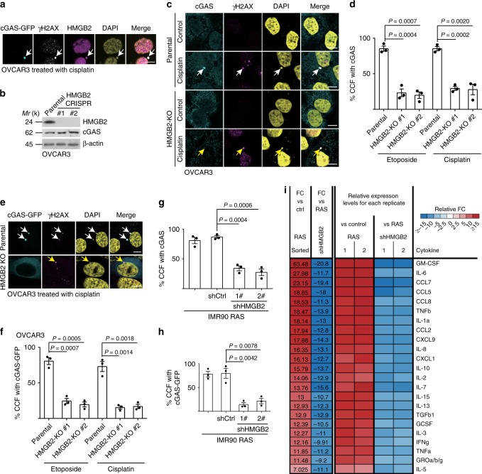
Fig. 1. HMGB2 is required for cGAS’ localization into CCF during senescence.
a OVCAR3 cells expressing GFP-tagged cGAS were induced to senesce by cisplatin, and imaged under a confocal microscope. cGAS-GFP, γH2AX, and HMGB2 co-localized CCF are indicated by arrows. b Expression of HMGB2, cGAS, and a loading control β-actin in the indicated parental and two independent HMGB2 knockout OVCAR3 clones determined by immunoblot. c, d Representative images (c) and quantification (d) of endogenous cGAS localization into γH2AX-positive CCF in senescent parental and HMGB2 knockout OVCAR3 cells with the indicated treatment. Arrows point to CCF. e, f Representative images (e) and quantification (f) of cGAS-GFP localization into γH2AX-positive CCF in senescent parental and HMGB2 knockout OVCAR3 cells with the indicated treatment. Arrows point to CCF. g, h Quantification of endogenous cGAS (g) or cGAS-GFP (h) localization into CCF in senescent primary IMR90 cells induced by oncogenic RAS, with or without HMGB2 knockdown. i The secretion of soluble factors under the indicated conditions were detected by antibody arrays. The heatmap indicates the fold change (FC) in comparison with the control (Ctrl) or RAS-induced senescent IMR90 cells. Relative expression levels per replicate and average fold change differences are shown. Data represent mean ± s.e.m. n = 3 biologically independent experiments unless otherwise stated. Scale bar = 10 μm. P-values were calculated using a two-tailed t test. Source data are provided as a Source Data file.

