. 2020 Feb 13;12(1):76–85. doi: 10.4055/cios.2020.12.1.76

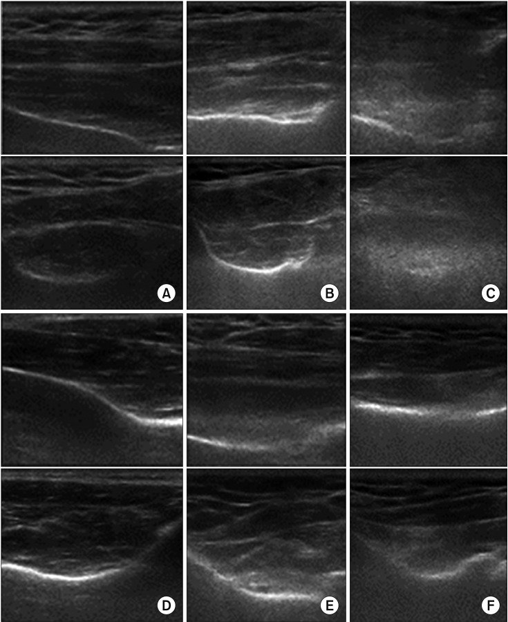
Fig. 2. Strobel's classification by ultrasonography. (A) Long- and short-axis views showing grade 0 echogenicity and grade 0 architecture of the supraspinatus muscle, which is isoechoic to the overlying muscle and has clearly visible intramuscular tendons and an identifiable pennate pattern. (B) Long- and short-axis views showing grade 1 echogenicity and grade 1 architecture of the supraspinatus muscle, which slightly increased echogenicity compared with the overlying muscle and partially visible intramuscular tendons and pennate pattern. (C) Long- and short-axis views showing grade 2 echogenicity and grade 2 architecture of the supraspinatus muscle, markedly increased echogenicity compared with the overlying muscle and no discernible intramuscular tendons or pennate pattern. (D) Long- and short-axis views showing grade 0 echogenicity and grade 0 architecture of the infraspinatus muscle, which is isoechoic to the overlying muscle and has clearly visible intramuscular tendons and an identifiable pennate pattern. (E) Long- and short-axis views showing grade 1 echogenicity and grade 1 architecture of the infraspinatus muscle, which slightly increased echogenicity compared with the overlying muscle and partially visible intramuscular tendons and pennate pattern. (F) Long- and short-axis views showing grade 2 echogenicity and grade 2 architecture of the infraspinatus muscle, markedly increased echogenicity compared with the overlying muscle and has no discernible intramuscular tendons or pennate pattern.

Fig. 2