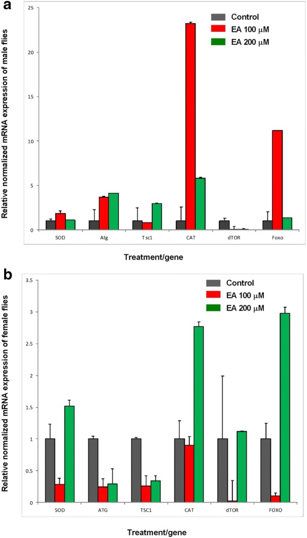
Fig. 4.

a Gene expression in male flies. Three vials were set up, with 30 flies/vial. Experiments were performed two times (n = 3 + 3). mRNA of manganese-containing superoxide dismutase (SOD2), autophagy gene (ATG1), tuberous sclerosis (TSC1), catalase (CAT), mammalian target of rapamycin (TOR), and forkhead box transcription factor (FOXO) in wild-type Canton-S male fruit flies fed with ellagic acid at the concentration of 100 μM and 200 μM. Rpl32 is used as internal control. Levels of gene expression in all groups were normalized with the control group for each gene. Error bars show standard deviation. b Gene expression in female flies. Three vials were set up, with 30 flies/vial. Experiments were performed two times (n = 3 + 3). mRNA of manganese-containing superoxide dismutase (SOD2), autophagy gene (ATG1), tuberous sclerosis (TSC1), catalase (CAT), mammalian target of rapamycin (TOR), and forkhead box transcription factor (FOXO) in wild-type Canton-S female fruit flies fed with ellagic acid at the concentration of 100 μM and 200 μM. RPL32 is used as internal control. Levels of gene expression in all groups were normalized with the control group for each gene. Error bars show standard deviation
