Figure 1.

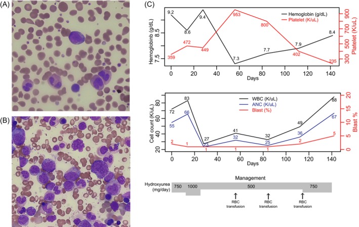
Peripheral blood smear (A) and bone marrow aspirate (B) in Wright and Giemsa stains. A blast and dysplastic granulocytes were shown (A) and granulocytic proliferation and granulocytic dysplasia in bone marrow (B). C, Laboratory findings including hemoglobin, platelet, white blood cell count, and blast count during the 140 days of hydroxyurea therapy
