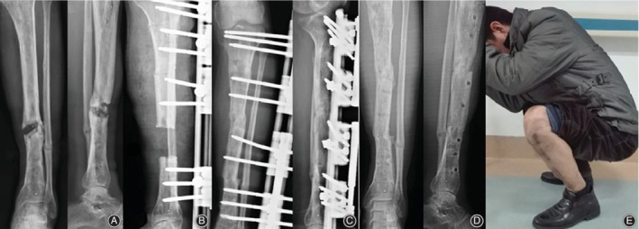
Figure 3.

An 42‐year‐old male patient with posttraumatic osteomyelitis the left tibia presented to our department and treated using LRS trifocal bone transport from proximal to distal. (A) Segmental defect of the left tibia caused by infection on X‐ray AP view. (B) Excision of infection bone with 7 cm defect and application of LRS with double level osteotomies for trifocal bone transport. (C) Bone transport was completed with good regenerate consolidation and docking union was achieved and evaluated on AP view of X‐ray at 4 months after index surgery. (D) LRS was removed with excellent bone result shown on AP view of X‐ray at 6 months after operation. (E) Functional recovery at last visit on squatting position at 34 months.
