Figure 4.

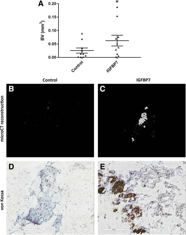
IGFBP7‐treated fibroblasts form mineralized tissue in vivo. Fibroblasts treated without or with IGFBP7 (1000 ng/mL) were implanted into nude mice using a carrier of Matrigel and ceramic microparticles. Mineralized tissue was analyzed by MicroCT to measure the total volume of mineralized bone tissue (BV) and von Kossa staining for detection of mineralization in tissue. MicroCT analysis of mineralized bone (0.2 g/cm3 threshold) showed a significant increase for IGFBP7‐treated fibroblasts (1 μg/mL) *P < .05 vs untreated controls (A). 3D reconstructed images from MicroCT analysis are shown (B,C). Images of histological sections stained with Von Kossa are shown (D,E). Scale bars shown in (D) and (E) indicate 300 μm
