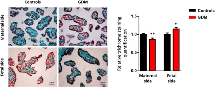
Figure 1.

Histological features of the placental tissue from maternal and fetal sides obtained from gestational diabetes mellitus (GDM) and control women. Representative photomicrographs of terminal and intermediate villi in placental sections from pregnant control and GDM women, stained with Masson's trichrome to highlight connective tissue and collagen fibers (in green). Quantification of Masson's trichrome staining in villous stroma of placental sections is shown as percentage mean area ± SD (n = 6‐8 per group). Results are shown as mean ± SEM from independent donor experiments performed in duplicate. *P < .05 vs controls, **P < .01 vs controls
