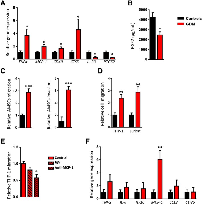
Figure 3.

Disturbances in amniotic mesenchymal stem cells (AMSCs) and amniotic membrane‐resident macrophages from placentas obtained from gestational diabetes mellitus (GDM) women. A, Gene expression analysis of the inflammatory markers TNFα, MCP‐1, CD40, CTSS, IL‐33, and PTGS2 in AMSCs isolated from pregnant control and GDM women (n = 8 per group). B, Prostaglandin E2 (PGE2) levels in the conditioned medium of AMSCs from pregnant control and GDM women (n = 6‐7 per group). C, Migratory and invasive capacities of AMSCs isolated from pregnant control and GDM women assessed in Transwell assays (n = 7‐9 per group). D, Migration of THP‐1 and Jurkat cells to AMSC‐conditioned medium assessed in Transwell assays (n = 5‐7 per group). E, Migration of THP‐1 cells to GDM‐AMSC‐conditioned medium after incubation with anti‐MCP‐1 antibody. F, Gene expression analysis of pro‐inflammatory and chemotactic markers (TNFα, IL‐6, IL‐1β, MCP‐1, CCL3, and CD86) in amniotic membrane‐resident macrophages from pregnant control and GDM women (n = 6‐8 per group). Results are shown as mean ± SEM from independent donor experiments performed in duplicate. *P < .05 vs controls, **P < .01 vs control, ***P < .0001 vs controls
