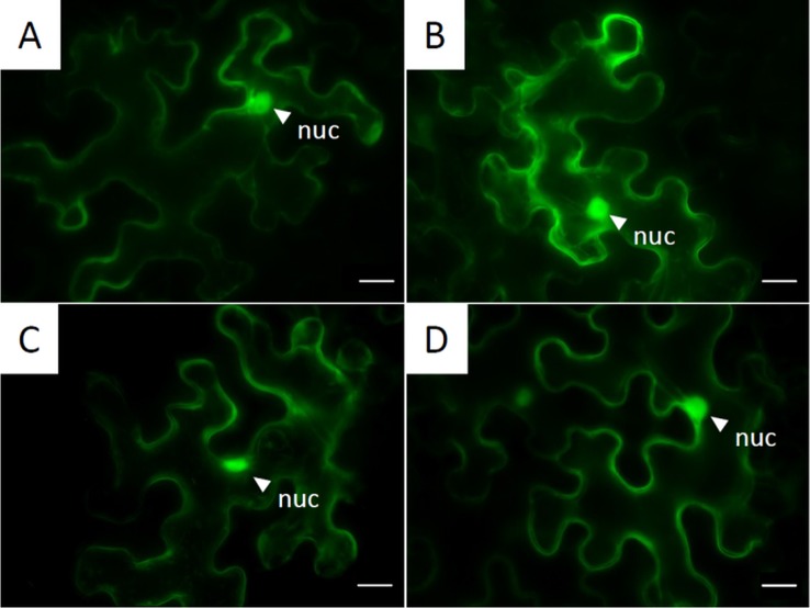
Figure 6.
Subcellular localization of GFP-tagged NLPs inside the cytoplasm: Pictures are obtained from fluorescent microscopy of transiently expressed C-terminal GFP-labeled NLPs in N. benthamiana 72 h post infiltration. (A) PvNLP1; (B) PvNLP2; (C) PvNLP3; (D): enhanced GFP (untagged); nuc, nucleus; scale bar = 20 µm.

