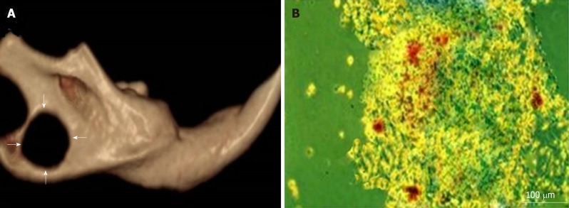
Figure 2.
CBCT and inverted microscopic imaging. A: CBCT images of critical-sized defects (arrows) in rats' mandibles; B: An inverted light microscopic image of bone marrow-derived stem cells differentiated in osteogenic medium with mineralized deposits identified by alizarin red staining on the 7th day.

