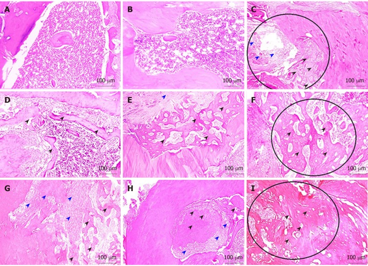
Figure 4.
Hematoxylin and eosin staining. A-C: Decalcified 4-µm thick sections showing the empty bone defect area of group I at 1, 2 and 4 wk; D-F: the bone defect area of group II grafted with rich fibrin membrane at 1, 2 and 4 wk; G-I: the bone defect area of group III grafted with platelet-rich fibrin membrane and seeded with bone marrow-derived stem cells at 1, 2 and 4 wk. Black arrowheads (osteoid bone); red arrowheads (granulation tissue).

