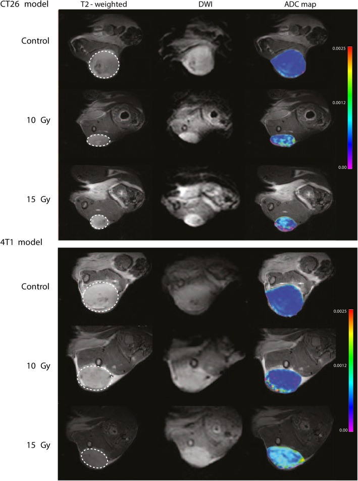
Fig. 2.
MRI images shown for both anatomical T2-weighted scan, DWI scan from shortest b-value, and overlay of anatomical image and ADC-map. Depicted is one mouse from each group in both models. The T2-weighted anatomical sequence was performed on Bruker 7 T preclinical MRI system using the following parameters; TR/TE. 2500/35 milliseconds, image size: 256 × 256, Field of view (FOV): 30 × 30 mm, averages: 2, slice thickness: 0.7 mm, and scan time 2 min 40 s. Diffusion-weighted scan sequence was performed using the following parameters; TR/TE: 550/24 milliseconds, image size: 96 × 96, FOV: 30 × 30 mm, averages: 6, segments: 6, slice thickness: 0.7 mm, b-values: 0, 100, 200, 600, 1000, 1500, 2000, and scan time 2 min 18 s

