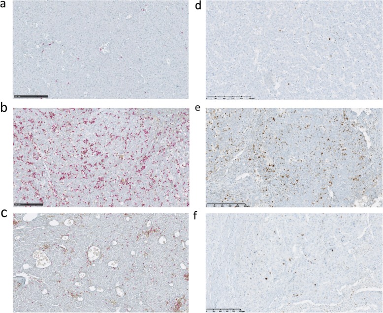
Fig. 1.
Representative images of dual CD4/ CD8 and Granzyme B staining on tumor tissues. Scans were imaged at 10x magnification using NDPview software (Hamamatsu). a TILs in a non-treated HCC patient, showing CD4+ (brown) and CD8+ (red) cells. b TILs in a preoperative SIRT-treated HCC patient, showing increased infiltrates with CD4+ (brown) and CD8+ (red) cells. c TILs expression in a preoperative TACE-treated HCC patient showing similar infiltrates with CD4+ (brown) and CD8+ (red) cells as observed in untreated patients but associated with significant areas of necrosis. d Granzyme B expression in a non-treated HCC patient (brown). e Granzyme B expression in a preoperative SIRT-treated HCC patient. f Granzyme B expression in a preoperative TACE-treated HCC patient

