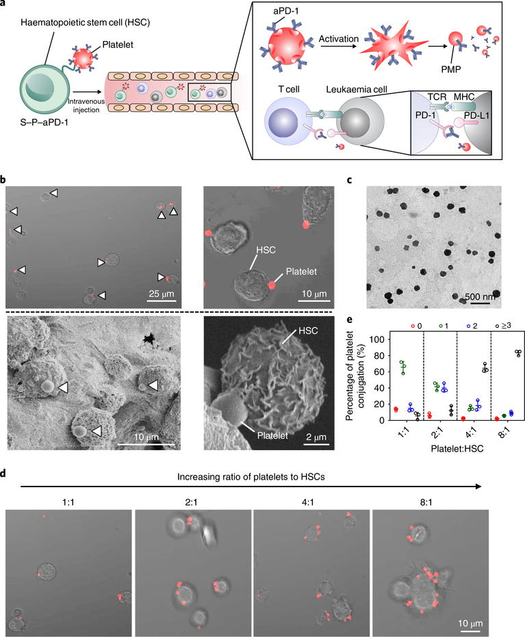
Fig. 1 |. Characterization of the S–P–aPD-1 cellular combination delivery system.
a, Schematic of HSC–platelet assembly-assisted aPD-1 delivery. After intravenous delivery, the S–P–aPD-1 could home to the bone marrow and the platelets could be locally activated and release aPD-1 to bind T cells for an enhanced immune response. MHC, major histocompatibility complex; TCR, T-cell receptor. b, Confocal microscopy (top) and SEM characterization (bottom) of S–P–aPD-1 conjugates. The platelets were labelled with rhodamine B for confocal observation. White arrows indicate the presence of platelets. c, Transmission electron microscopy (TEM) characterization of PMPs from S–P–aPD-1 after activation by 0.5 U ml−1 thrombin. d, Fluorescence imaging of S–P–aPD-1 with different ratios of platelets to HSCs. e, Quantitative analysis of conjugated platelets on HSCs (the number of bound platelets per cell is indicated as 0, 1, 2, 3.). The quantification was based on counting HSCs (numbers = 200) under the confocal microscope. The experiments were repeated three times. Data are presented as means ± s.d.

