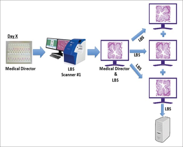
Figure 7.
Intra- and inter-pathologist precision study: Scanning and FOV preparation schema. The entire set of 69 microscope slides was scanned once at Site #1 using a single scanner. For each feature, a FOV was extracted and saved in three different orientations. FOV: Fields of view. LBS: Leica Biosystems

