Fig. 2.

Scanning electron micrographs of (A) the SA gel beads and (B) the CS-SA gel beads, the scale bar was 0.25 mm and 50 μm, respectively.

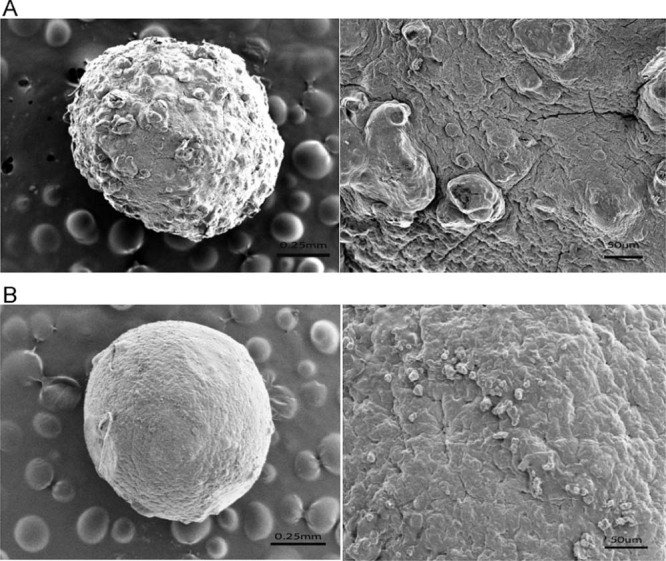
Scanning electron micrographs of (A) the SA gel beads and (B) the CS-SA gel beads, the scale bar was 0.25 mm and 50 μm, respectively.