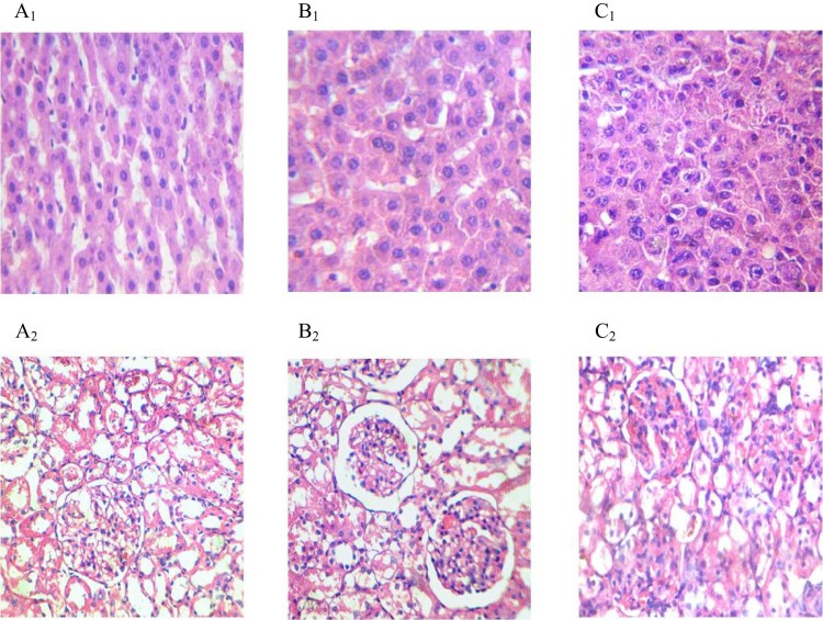
Fig. 4.
Histological alterations in the liver and kidney at 21 d after administration of pure HP and Au-mPEG(5000)-S-HP nanoparticles (H&E staining, 40×). Both (A1) and (A2) photomicrograph of liver and kidney section of control rat demonstrated normal cellular structure. Both (B1) and (B2) sections of liver and kidney of pure HP drug at 20 mg/kg b.wt show normal cellular architecture and both (C1) and (C2) section of liver and kidney of Au-mPEG(5000)-S-HP nanoparticles drug at 0.5 ml depicted normal cellular architecture.

