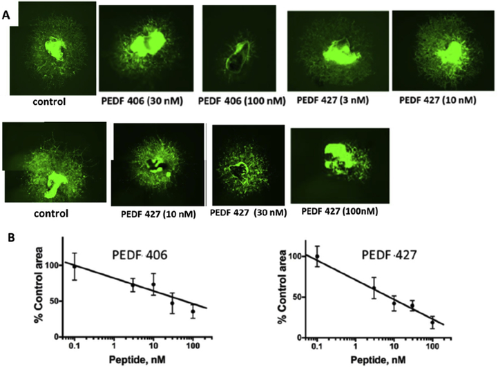
Figure 2. PEDF peptides inhibit choroidal neovascularization ex vivo.
Choroidal explants were generated from eyes harvested from C57Bl6 mice that carry eGFP transgene under beta-actin promoter. The explants were embedded in Matrigel drops and cultured in media containing VEGF alone or with increasing concentrations of selected PEDF peptides from Table 1. The explants were observed daily and images were acquired at 6 days endpoint. Composite images were generated for larger vascular areas to evaluate the full vascular area. (A) Representative images of VEGF-stimulated choroidal explants treated with PEDF 406 at increasing concentrations. Similar images were obtained for PEDF 427. (B) Quantitative analysis of the images as shown in A. Sprouting area was measured using ImageJ software and the area of explant subtracted for each image. A minimum of 3 explants were analyzed per data point. Statistical significance was determined using multiple T-test and P value is below 0.0001 for both C and D.

