Figure 2.

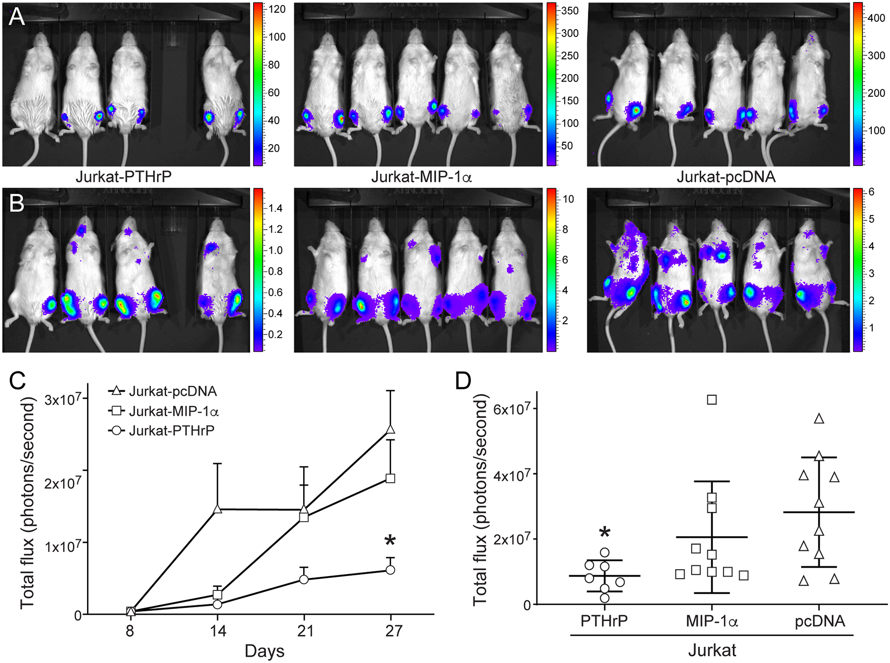
In vivo bioluminescent imaging showing Jurkat-PTHrP tibial neoplasms grew at a significantly slower rate than JurkatpcDNA neoplasms. Bioluminescent imaging of mice with Jurkat cells with overexpression of PTHrP, MIP-1α, or vector mRNA control. (A) Successful tibial engraftment was confirmed on the first week of bioluminescent imaging. Y-axes are × 103. (B) On day 30 (sacrifice) tibial tumor burden had increased in all groups. Y-axes are × 106. (C) The Jurkat-pcDNA tibial neoplasms had the greatest bioluminescent signal. Jurkat-pcDNA bone neoplasms grew at a significantly faster rate than Jurkat-PTHrP bone neoplasms (*p <.05). (D) On the day of sacrifice, overall tibial tumor burden was significantly decreased in the Jurkat-PTHrP mice, as determined by decreased intensity of bioluminescence in the tibias (*p < .05).
