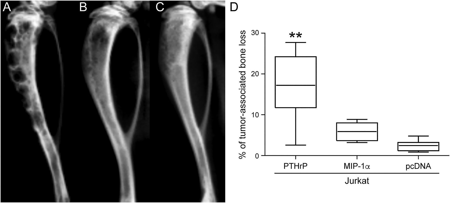
Figure 3.

Radiographic bone loss was significantly greater in the Jurkat-PTHrP tumor-bearing tibias. (A) Tibias with Jurkat-PTHrP neoplasms had significantly greater bone loss as indicated by the total area of radiolucency. Multifocal to coalescing areas of radiolucency spanned from the proximal metaphysis to the mid- to distal diaphysis in Jurkat-PTHrP-bearing bone neoplasms. (B) Jurkat-MIP-1α -bearing tibias had focal to coalescing areas of radiolucency that were significantly less than the Jurkat-PTHrP neoplasms but more than Jurkat-pcDNA tumor-bearing tibias. (C) Jurkat-pcDNA tibial bone neoplasms had minimal areas of radiolucency. (D) Bone loss was quantified on high-resolution faxitron images using Bioquant software. Jurkat-PTHrP mice had significantly greater tumor-associated bone loss than Jurkat-MIP-1α and JurkatpcDNA (**p < .01).
