Figure 4.

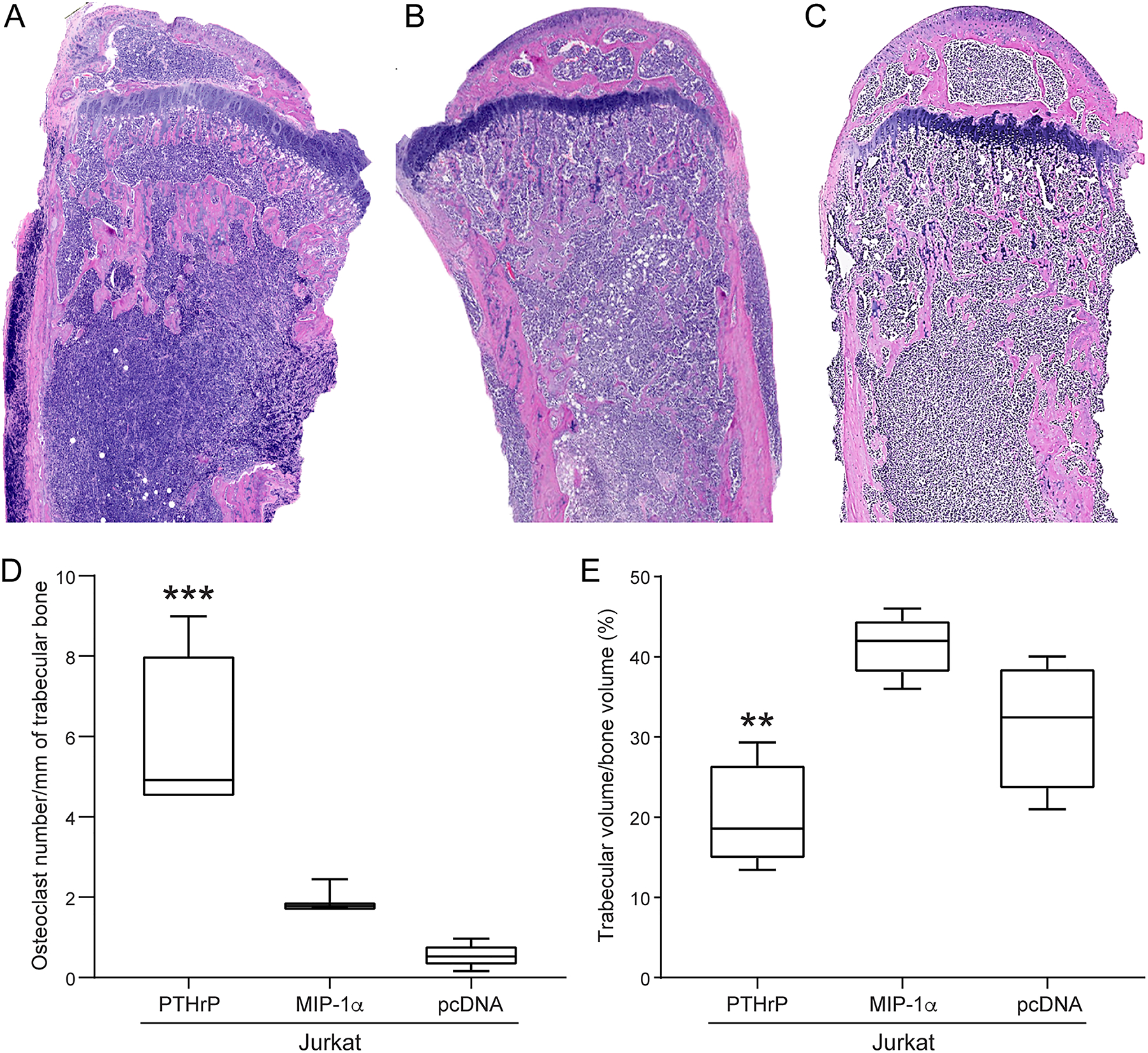
Jurkat-PTHrP neoplasms had increased osteoclast number and decreased trabecular bone volume. (A) H&E-stained tibias from the Jurkat-PTHrP mice had lymphoma cells that spanned from the proximal metaphysis to the mid- to distal diaphysis. A decrease in the number and total area of trabecular bone was present. Tibias of Jurkat-MIP-1α (B) and Jurkat-pcDNA (C) mice had lymphoma cells spanning from the metaphysis to varying depths of the tibial diaphysis. Endosteal new bone formation was present adjacent to the lymphoma cells within the metaphysis in both Jurkat-MIP-1a and Jurkat-pcDNA mice. (D) The number of osteoclasts per surface of trabecular bone and the number of osteoclasts was significantly increased in Jurkat-PTHrP tumor-bearing tibias when compared with Jurkat-pcDNA and Jurkat-MIP-1α mice (***p < .001). (E) The area of trabecular bone was significantly decreased in the Jurkat-PTHrP mice compared to the Jurkat-pcDNA and Jurkat-MIP-1α mice (**p < .004).
