Figure 5.

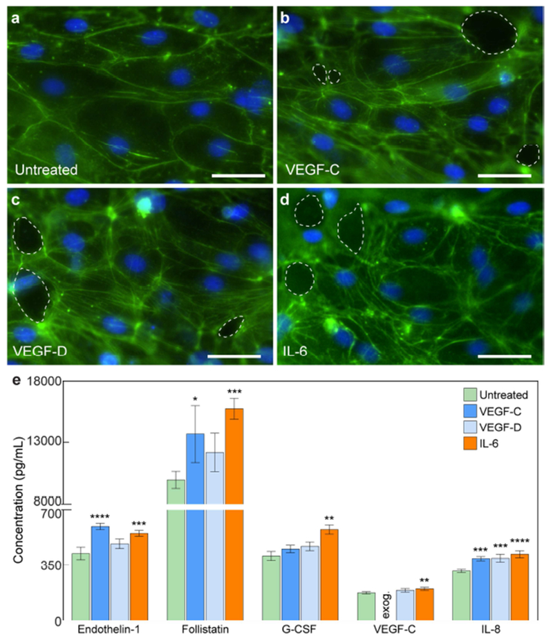
Morphological and secretion responses of lymphatic vessels to exogenous lymphangiogenic and inflammatory stimuli. a-d Images of lymphatic endothelia under VEGF-C, VEGF-D, and IL-6 stimulation (F-actin in green and nuclei in blue). In comparison to untreated vessels, LECs in stimulated vessels express increased actin stress fibers. There are also holes the vessel wall (dashed outlines). e Cytokine concentrations for untreated and stimulated conditions. VEGF-C and IL-6 induce significant changes in all presented cytokines. Concentration values are the averages of n = 4 technical replicates of media pooled from 6 individual vessels for each condition. All error bars are one standard deviation. Scale bars are 10 μm. *p ≤ 0.05, **p ≤ 0.01, ***p ≤ 0.001, ****p ≤ 0.0001.
