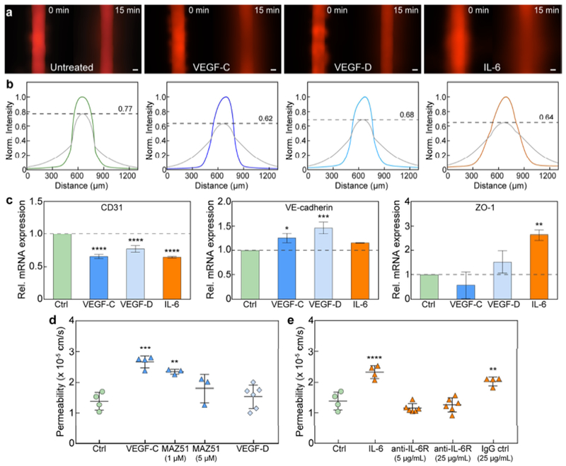
Figure 6.

Barrier response of lymphatic vessels to exogenous lymphangiogenic and inflammatory stimuli. a Images of 70 kDa dextran diffusion in untreated vessels and vessels stimulated with VEGF-C, VEGF-D, and IL-6. b Normalized intensity profiles of dextran diffusion. The initial concentration decreases by 38%, 32%, and 36% for VEGF-C, VEGF-D, and IL-6 stimulation, respectively, as compared to 23% for untreated vessels. c Transcriptional expression of intercellular junctions under stimulation. CD31 expression is significantly downregulated for all cases. VE-cadherin expression is upregulated after VEGF-C and VEGF-D treatment, and ZO-1 after IL-6 stimulation. Relative mRNA values are the averages of n = 3 technical replicates with each replicate representing two individual vessels. d Vessel permeability significantly increases after VEGF-C treatment. Barrier function can be rescued by inhibiting the phosphorylation of VEGFR3, the receptor for VEGF-C, with MAZ51 (partially at 1 μM and fully at 5 μM). e IL-6 stimulation significantly reduces barrier capacity, which can be mitigated via antibody-mediated blocking of the IL-6 receptor on the lymphatic vessels. All permeability values are the averages of at least n = 3 individual vessels. All error bars are one standard deviation. Scale bars are 100 μm. *p ≤ 0.05, **p ≤ 0.01, ***p ≤ 0.001, ****p ≤ 0.0001.
