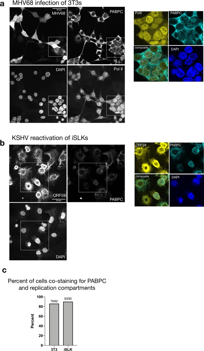
Fig 5. PABPC is not excluded from replication compartments.
(A) MHV68 infected 3T3 cells were subjected to imunofluoresence (IF) analysis at 27 hpi using antibodies against PABPC and Pol II, and stained with DAPI. The MHV68 genome contains GFP, which served as a marker of infection. Cells with RCs are outlined in white. RCs were identified in cell nuclei as regions that contained Pol II but stained poorly for DAPI. The inset shows a merge of Pol II and PABPC staining for several cells that co-stain for both proteins in RCs. (B) IF was performed on KSHV-positive iSLK cells reactivated for 48 h and stained with antibodies against PABPC, ORF59 and DAPI. Cells with RCs are outlined in white and were identified as nuclei with ORF59 staining that overlaps with regions that stain poorly with DAPI. The inset shows a merge of ORF59 and PABPC for several cells that co-stain for both proteins in RCs. (C) Percent of RC containing cells that overlap with PABPC signal. Fractions of cells with PABPC signal in RCs over total number of cells counted with RCs are displayed.

