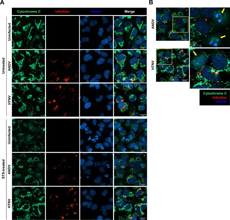
Fig 2. Hantavirus infection inhibits STS-mediated release of cytochrome C.
A549 cells were infected at MOI 1 and treated with STS for 4 hours at 72 hours p.i. (A) Immunofluorescence images showing cytochrome C (green) in ANDV- and HTNV-infected cells (red) after exposure to STS. DAPI (blue) was used for nuclear counterstaining. One representative experiment out of three independent experiments is shown. Scale bar, 20 μm. (B) Magnified immunofluorescence pictures showing cytochrome C (green) release in bystander uninfected cells (yellow arrows) but not in ANDV- and HTNV-infected cells (red). DAPI (blue) was used for nuclear counterstaining. Yellow arrows indicate bystander uninfected cells. One representative experiment out of three independent experiments is shown. Scale bar, 20 μm.

