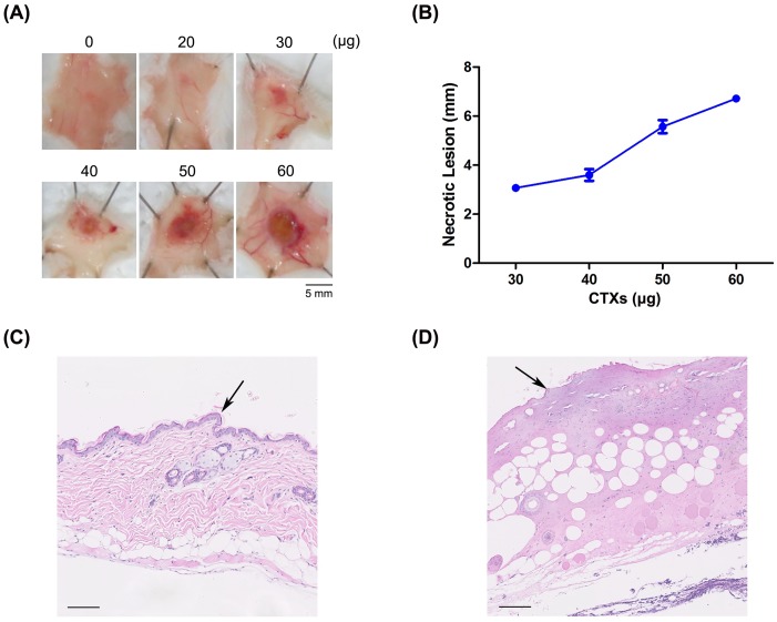
Fig 4. CTX-induced necrosis in mouse models.
(A) Different amounts (0–60 μg) of CTXs were intradermally injected into mice, and the sizes of necrotic lesions in the dorsal skin of injected mice were measured. (B) MNDs for CTXs, calculated from measured sizes of necrotic lesions. H&E-stained sections of dorsal skins injected with (C) saline solution or (D) CTXs were analyzed histologically under a light microscope. Arrows highlight alterations in the epidermis. Scale bar: 100 μm.

