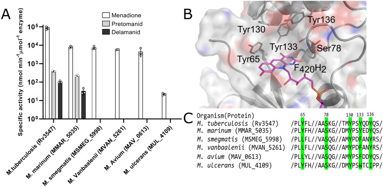
Fig 2. The activity of Ddn and orthologs from other mycobacterial species and key substrate binding pocket residues.
(a) The activity of Ddn and orthologs with menadione, pretomanid, and delamanid. Error bars show standard deviations from three independent replicates. (b) Multiple sequence alignment of Ddn and orthologs. Highlighted residues indicate active site residues and numbers indicate their residue position in Ddn. (c) The binding pocket of Ddn consists of F420/F420H2, Tyr65, Ser78, Tyr130 and Tyr136.

