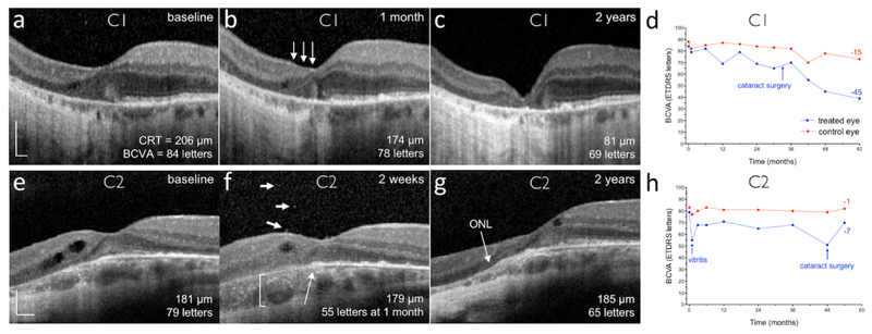
Figure 3. Retinal structural and visual acuity changes in participants C1 and C2.
Subretinal delivery of gene therapy vector in participant C1 (a-d) was complicated by retinal stretch, resulting in a reduced vector dose: spectral-domain optical coherence tomography (OCT) cross-section through the fovea of the treated (left) eye at (a) baseline, (b) 1 month and (c) 2 years. One month after surgery, a reduction in outer nuclear layer (ONL) thickness was noted nasal to the fovea (arrows), although the temporal half of the fovea and the maximal retinal thickness remain similar to baseline. By 2 years, retinal thinning had stabilised, but the best-corrected visual acuity (BCVA, number of ETDRS letters) has reduced consistent with the foveal collapse (d). CRT, central retinal thickness. (e-h) OCT cross-section through the fovea of the treated (left) eye at (e) baseline, (f) 2 weeks (note visual acuity is from the 1 month visit) and (g) 2 years in participant C2, who experienced significant intraocular inflammation after gene therapy. This patient did not have a clear ellipsoid zone layer before surgery and subtle cystic degenerative changes can be seen nasally. Two weeks after gene therapy, vitreous cells (f: short arrows), outer retinal opacities (f: long arrow) and choroidal thickening (f: bracket) could be seen around the fovea. This was associated with an acute drop in BCVA, which recovered partially after a prolonged course of oral corticosteroid (h). The clear laminated appearance of the ONL appears to have improved by 2 years (g). Scale bars represent 200 μm.

