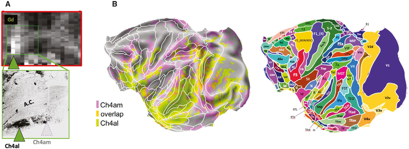
FIGURE 7.
Disruption of subcortical neural activity alters cortical functional magnetic resonance imaging (fMRI) patterns. Transient pharmacological inactivation of two subregions in the nucleus basalis, Ch4al and Ch4am (A), induced spatially distinct changes in the amplitude of cortical fMRI signals (B) in the macaque. For anatomic reference, superimposed on the map in B are areal boundaries of the Saleem and Logothetis atlas. Adapted with permission from Turchi et al73

