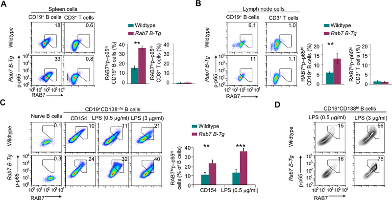
FIGURE 4.
RAB7 potentiates NF-κB activation. (A,B) Flow cytometry analysis of intracellular levels of RAB7 expression and NF-κB p65 phosphorylation in B and T cells in the spleen (A) and pooled lymph nodes (B) of immunized mice. Numbers indicate the proportions of B cells showing high RAB7 expression and p65 phosphorylation (mean and s.e.m., n=3 in the histograms). (C) Flow cytometry analysis of RAB7 expression and p65 phosphorylation in freshly isolated naïve B cells and B cells stimulated in vitro with CD154 or LPS for 48 h (mean and s.e.m., n=4). (D) Flow cytometry analysis of RAB7 expression and p65 phosphorylation in plasma cells generated after in vitro B cell stimulation by LPS, as indicated, for 48 h (CD154 stimulation did not generate enough plasma cells for analysis). Representative of three independent experiments. **, p < 0.01; ***, p < 0.005.

