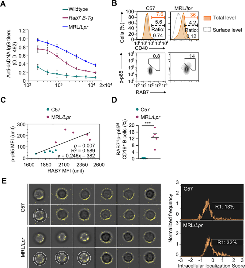
FIGURE 7.
CD40 intracellular localization in lupus B cells. (A) ELISA of titers of circulating anti-dsDNA autoantibodies in Rab7 B-Tg mice and their wildtype counterparts (n=3; mean and s.d.). (B) Flow cytometry analysis of total (orange shaded area) and surface (dashed black lines) CD40 expression in B cells from PBMCs of MRL/Lpr and C57 mice (top panels). The ratio of cells displaying only surface CD40 expression, as the readout of the inverse of CD40 internalization. Also depicted is flow cytometry analysis of B cells showing high RAB7 expression and NF-κB p65 phosphorylation in such B cells (bottom panels (bottom panels). (C) Linear regression of intracellular RAB7 expression and levels of phosphorylated p65 in B cells from PBMCs of MRL/Lpr and C57 mice, as determined by MFIs in flow cytometry (right; n=5; p value, F test). (D) RAB7 expression and p65 phosphorylation in B cells from PBMCs of MRL/Lpr and C57 mice (mean and s.d., n=5 in the histogram). (E) Flow imaging analysis of CD40 intracellular localization in CD19+ spleen B cells. The images of B cells showing CD40 intracellular localization, i.e., 1 out of 11 representative C57 B cells and 3 out of 9 representative MRL/Lpr B cells, were duplicated beneath for the annotation by white concentric circles of the peripheral dark ring, which indicated the cell surface and resulted from lack of surface staining of CD40. Also depicted is the proportion of B cells showing CD40 intracellular localization score of >0 (histogram, right). B, E, representative of at least two independent experiments). ***, p < 0.005.

