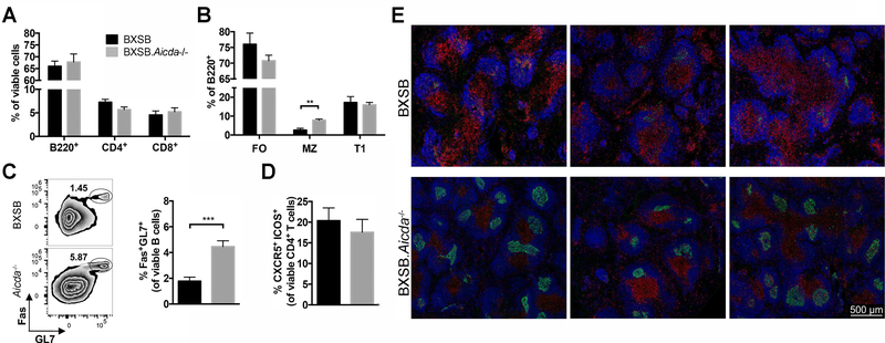
FIGURE 4.
Marginal zone B cell depletion and germinal center integrity restored in BXSB.Aicda−/− mice. Splenocytes of 12-week-old BXSB (n=7) and BXSB.Aicda−/− (n=9) males were analyzed. (A) The percentages of B220+, CD4+ and CD8+ cells in the viable cell population were compared between wt and Aicda−/− mice. (B) The percentages of follicular (FO, CD23+ CD21+), marginal zone (MZ, CD23low/- CD21+) and transitional 1 (T1, CD23- CD21-) B cells in the B lymphocyte population are shown. (C) Representative flow cytometric zebra plots showing the percentage of germinal center (GC) B cells (Fas+ GL7+) in the viable B lymphocyte population with summary data shown on the right. (D) The percentage of TFH cells (CXCR5+ ICOS+). (E) Representative confocal images showing the follicles in the spleen. Blue: B220, Green: GL7 and Red: CD4. Data are presented as mean±SEM. Statistical significance was determined by Mann-Whitney U test. **p<0.01, ***p<0.001. Results are representative for at least 3 independent experiments.

