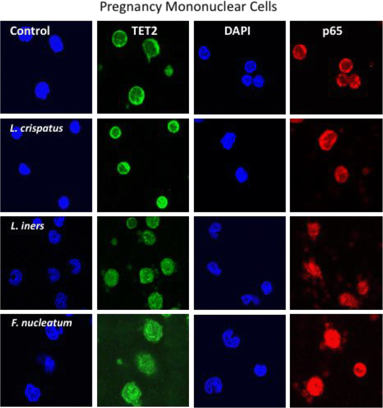
Fig. 3.
Representative images of mononuclear cells isolated from normal pregnant women treated with commensal bacteria. Green fluorescence identifies TET2, red identifies the p65 subunit of NF-κB, and DAPI blue identifies the nucleus. In untreated cells, both TET2 and p65 were primarily localized to the cytosol. L. crispatus, L. iners, and F. nucleatum all increased the translocation of TET2 and p65 from the cytosol to the nucleus, with F. nucleatum causing the most intense nuclear localization. The dose for bacteria was 100:1 (bacteria to cells)

