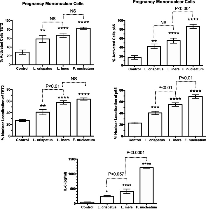
Fig. 4.
Percent of pregnancy mononuclear cells activated, % nuclear localization for TET2 and p65, and media concentrations of IL-8 in response to treatments with bacteria. Nuclear localization was quantitated by the density of immunofluorescence in the area of the nucleus as a percentage of the density of the entire cell using ImageJ software. Percentage of cells activated was quantified by counting the number of cells with brighter fluorescence in the nucleus than in the cytosol and taking that as a % of the total cells. The average number of cells counted per field was 27. L. crispatus, L. iners, and F. nucleatum all activated the cells causing translocation of TET2 and p65 from the cytosol to the nucleus with a parallel increase in IL-8 in a general order of L. crispatus < L. iners < F. nucleatum. (n = 8 for TET2, n = 10 for p65, n = 11 for IL-8, data represent mean ± SE, *P < 0.05, **P < 0.01, ***P < 0.001, ****P < 0.0001 as compared with control. P values are for comparisons between bacterial treatments. NS, non-significant)

