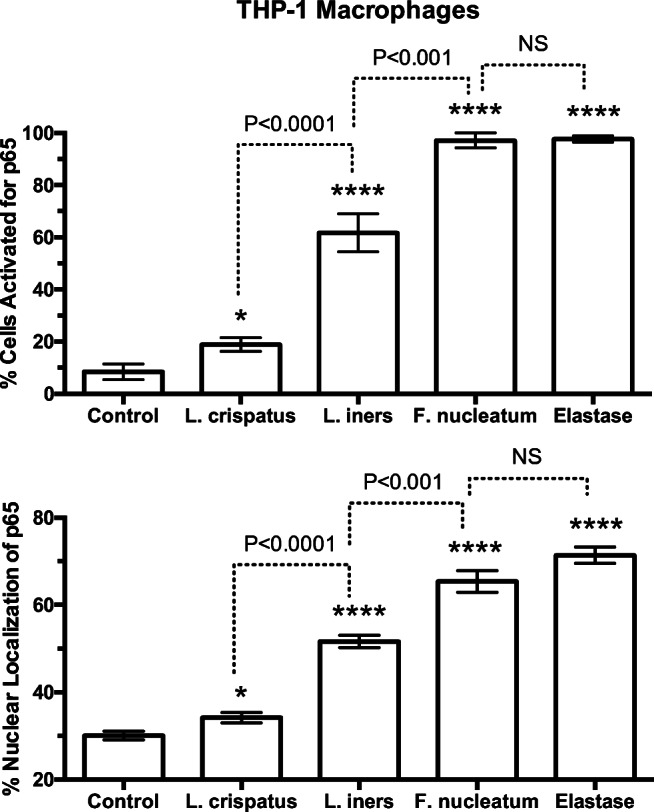
Fig. 8.

Percent of cells activated and % of nuclear fluorescence for the p65 subunit of NF-κB in THP-1 cells in response to treatments with bacteria or elastase. Percent of cells activated and % nuclear localization was quantitated as described for Fig. 4. The average number of cells counted per field was 20. L. crispatus activated 15% of cells; however, L. iners activated over 60% of cells and F. nucleatum activated almost all cells similar to protease treatment with elastase. (n = 5 for bacteria, n = 3 for elastase, mean ± SE, *P < 0.05, ****P < 0.0001 as compared with control. P values are for comparisons between bacterial treatments. NS, non-significant)
