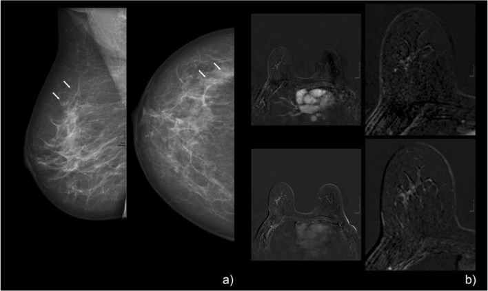
Fig. 5.
A 53-year-old woman with screen-detected calcifications in the right breast. Mammographic CC and MLO views (a) showed grouped, fine pleomorphic calcifications that were assigned BI-RADS 4b. Pre-interventional breast MRI (b) demonstrated a corresponding regional, heterogeneous, enhancing non-mass lesion in the right breast. Persistent enhancement kinetics (b: upper right, early enhanced; lower right, late enhanced subtraction) and irregular margins resulted in a Kaiser score of 3 (corresponding to BI-RADS 2/3, benign finding). Stereotactic biopsy revealed non-specific proliferative changes in the tumor-free breast parenchyma with calcifications: B2. MRI follow-up (not shown here) showed no suspicious findings

