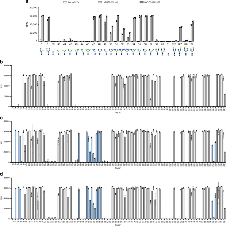
Fig. 2. Activity assay of HsFUT8 on glycan microarray.
a Aleuria aurantia lectin (AAL-555) binding before and after incubation with recombinant HsFUT8 at two different concentrations (650 nM and 3.5 μM) on a selection of glycan structures. b AAL-555-binding profile on a full glycan array before treatment with recombinant HsFUT8. c AAL-555-binding assay after treatment with recombinant HsFUT8 at 650 nM enzyme concentration and d AAL-555-binding assay after treatment with recombinant HsFUT8 at 3.25 μM enzyme concentration. Histogram bars show the average fluorescence RFU (relative fluorescence units) values for four replicate spots on the array. Error bars represent the standard deviation of the average RFU values on the same microarray. N-glycans that show binding towards AAL after incubation with HsFUT8 are highlighted in blue.

