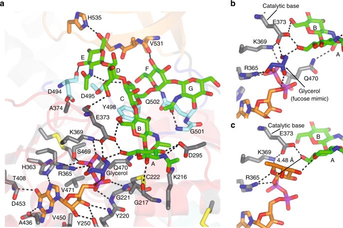
Fig. 5. Structural features of GDP-Fuc and G0 binding sites.
a Complete GDP-Fuc and G0 binding sites of the HsFUT8-GDP-G0 complex. The residues forming the GDP-Fuc/AG0/BG0 and CG0/DG0/EG0/FG0/GG0 binding sites are depicted as gray and aquamarine/orange carbon atoms, respectively. GDP, G0 and glycerol are shown as orange, green and blue carbon atoms, respectively. Hydrogen bond interactions are shown as dotted black lines. b, c Close-up view of the binding site region of the HsFUT8-GDP-G0 and HsFUT8-GDP-Fuc-G0 complexes showing the essential residues (Arg365, Lys369 and Glu373) that are major players in the plausible SN2 single-displacement reaction mechanism. Note the proximity and the orientation of the AG0 OH6 to the anomeric carbon (4.48 Å) which is compatible with the inversion of the configuration during the reaction.

