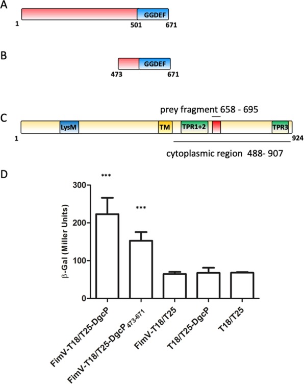
Figure 1.

DgcP interacts with FimV. Schematic diagrams of DgcP (A), its C-terminus cloned in the T25-DgcP473–671 construct (B) and FimV (C). The GGDEF domain of DgcP is shown, as well as the transmembrane region (TM), TPR motifs and the LysM domain of FimV. The red box in FimV corresponds to the prey fragment and lines show the regions cloned to confirm the interaction. The E. coli host strain BTH101 was co-transformed with pKT25_DgcP (full length) and pUT18_FimV constructs, with the T18 tag in the C-terminal; the interactions were measured using β-galactosidase activity as a reporter (C). Data are the means ± SD from three replicates. ***p < 0.001.
