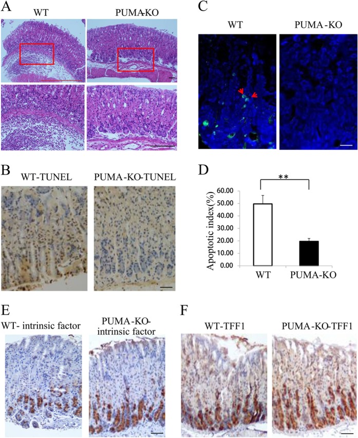
Fig. 3. Suppression of H. pylori infection-mediated carcinogenesis, apoptosis, and differentiation in PUMA-deficient mice (WT and PUMA-KO mice were treated with H. pylori for 2 months to induce gastritis).
a H&E staining of gastric tissues from WT and PUMA-KO mice after H. pylori treatment for 2 months (×100). Boxed regions of the glands are magnified in the lower panel (×200). b TUNEL IHC staining of gastric tissues from the treated mice (×200). c TUNEL (green) IF staining of gastric tissues from the treated mice after H. pylori treatment for 2 months, N = 5/group; the results are presented as the mean ± SEM. d The apoptosis index was measured by counting TUNEL signals in 100 randomly selected glands. **P < 0.01. e Staining for intrinsic factor showed mature zymogenic cells in H. pylori-treated wild type (left) and PUMA-KO (right) mice for 2 months. f Staining for TFF1 showed surface mucous cells in wild type (left) and PUMA-KO (right) mice treated with H. pylori for 2 months.

